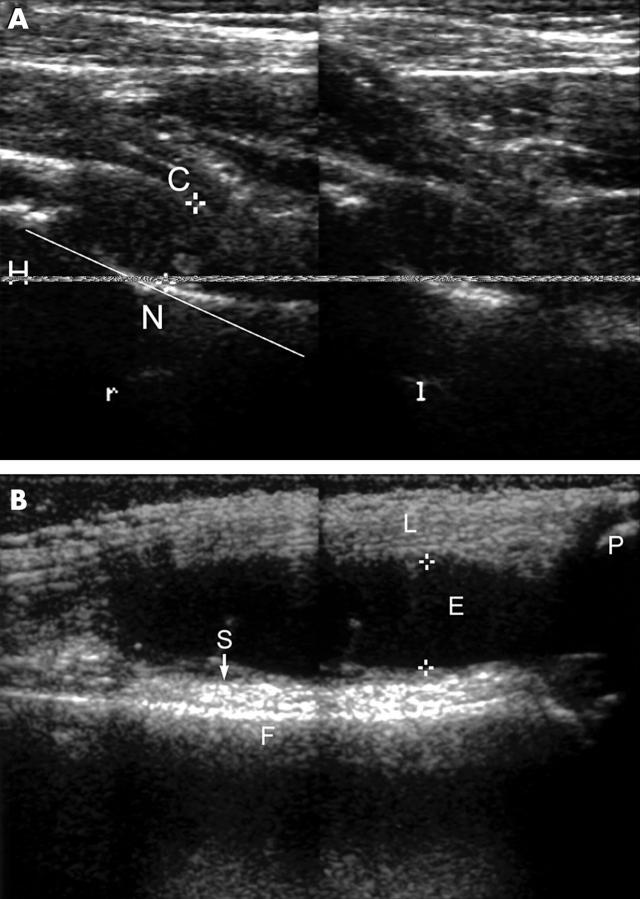
Figure 1.
Arthrosonography of the hip and knee joint. (A) Ultrasound of the hip joint in a ventral sagittal approach with the hip in extension, r = right and l = left side of the patient with active hip involvement in JRA. The scan shows the femoral head (H), the femoral neck (N), and the joint capsule (C). The distance between the femoral neck and the capsule is measured as the so-called synovial joint space (SJS). (B) Ultrasound of the knee joint in a ventral longitudinal scan of the suprapatellar pouch. Femur (F), patella (P), and the ligament (L) of the quadriceps are bordering the suprapatellar pouch. The joint effusion is measured in the largest anteroposterior diameter of the suprapatellar effusion (asterisks). In addition the maximum thickness of the synovial membrane (S) of the anterior wall is determined.

