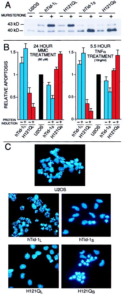
Figure 3.
hTid-1L and hTid-1S regulate apoptosis induced by mitomycin c and TNFα. (A) U2OS cells that express hTid-1L, hTid-1S, or J domain mutants (H121QL and H121QS, respectively) from a muristerone-inducible promoter were treated with muristerone for 24 hours (+) or went untreated (−) and were analyzed by immunoblot for the presence of hTid-1 proteins. (B) U2OS cells which express hTid-1L, hTid-1S or J domain mutants from a muristerone inducible promoter were either treated with muristerone (+) or mock-treated (−) for 24 hours and treated with 60 μM MMC for 24 hours (Left), or 10 ng/ml TNFα plus 30 μg/ml cycloheximide for 5.5 hours (Right), fixed, and stained with Hoechst. Apoptotic nuclei were counted and the numbers were compared with control cells. Rates of apoptosis in U2OS cells ranged from 20 to 30% for cells treated with MMC, and from 40 to 50% for cells treated with TNFα. The average of at least three independent experiments is shown. Error bars are ± 1 SD. (C) Fluorescence micrographs of Hoechst-stained U20S cells that inducibly express the indicated protein after 24-hour treatment with 60 μM MMC. Apoptotic cells display condensed and fragmented chromatin.

