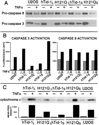
Figure 4.
hTid-1L and hTid-1S affect the rates of caspase 3 activation and cytochrome c release but not the rate of caspase 8 activation. Inducible U2OS cells expressing hTid-1L, hTid-1S, or J domain mutants (H121QL and H121QS, respectively) were treated with 10 ng/ml TNFα and cycloheximide for 4.5 hours (+) or went untreated (−). (A) Whole-cell lysates were analyzed by immunoblot for pro-caspase 8 and pro-caspase 3. (B) Lysates were analyzed for ability to cleave fluorogenic caspase 8 (IETD-AFC) or caspase 3 substrates (DEVD-AFC). (C) Cells were suspended in sucrose buffer and homogenized. Samples were centrifuged at 10,000 × g, and cytoplasmic extracts were analyzed by immunoblot for the presence of cytochrome c. Mean pixel densities of cytochrome c Western blot analysis are shown Lower.

