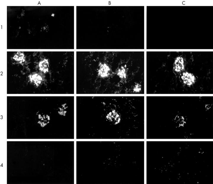
Figure 3 .
Immunohistology of kidney sections obtained from (NZBxNZW)F1 female mice. Kidney sections were obtained from 2 month old control mice (1A, 1B, 1C), 6 month old untreated mice (2A, 2B, 2C) and 6 month old tamoxifen treated (3A, 3B, 3C, 4A, 4B, 4C) (NZBxNZW)F1 mice. Mice were killed and their kidney sections (5 µm) were stained with FITC labelled goat antimouse IgG (total IgG; panel A), IgG2a (panel B), or IgG3 (panel C) using γ, γ2a, or γ3 heavy chain specific antibodies, respectively. Representative figures (x200) are presented. Sections 1 and 4 were defined as negative (score 0), section 2 as strongly positive (+++) and section 3 as positive (+/++). Note the relatively low intensity of IgG3 deposits in kidney sections obtained from tamoxifen treated mice (3C).

