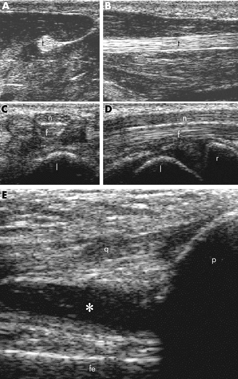
Figure 1.

(A, B) Healthy subject. Thenar eminence. Deep flexor tendon of the first finger (t). Transverse (A) and longitudinal (B) volar scans. (C, D) Healthy subject. Carpal tunnel. Median nerve (n) and finger flexor tendons (f). Transverse (C) and longitudinal (D) volar scans. l = lunate bone; r = radius. (E) Rheumatoid arthritis. Knee joint. Longitudinal suprapatellar scan. Suprapatellar pouch enlargement due to knee synovitis. * = synovial fluid; q = quadriceps tendon; p = patella; fe = femur.
