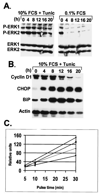
Figure 4.
Tunicamycin does not inhibit mitogenic signaling but blocks cyclin D1 translation. (A) After treatment of NIH 3T3 cells with tunicamycin or with medium containing 0.1% FCS, the levels of phosphorylated ERKs (Upper) were compared with the total ERK pool (Lower) by immunoblotting with antibodies that detect phosphorylated or all ERK isoforms, respectively. (B) After tunicamycin treatment of NIH 3T3 cells for the same time intervals, mRNAs encoding cyclin D1, CHOP, BiP, or actin were detected by Northern blotting performed with the cognate radiolabeled cDNA probes. (C) NIH 3T3 cells were left untreated (diamonds) or treated with 0.5 μg/ml tunicamycin for 2 h (squares), 4 h (triangles), or 12 h (circles) and then pulse-labeled for the indicated periods of time with Tran-35S-label (ICN). Cyclin D1 was immunoprecipitated from lysates, separated on a denaturing gel, and the rate of cyclin D1 synthesis was quantified by scanning the autoradiographs.

