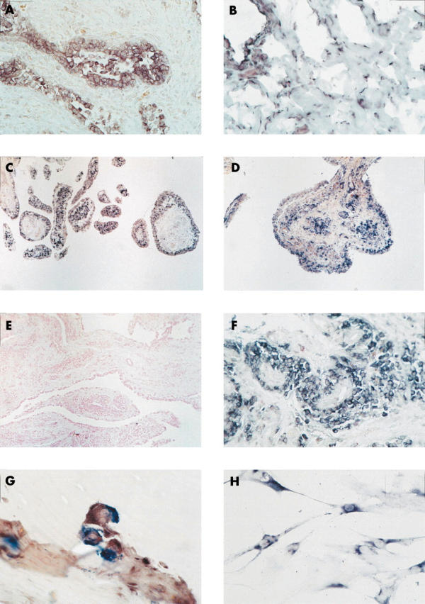
Figure 3.

In situ hybridisation on paraffin embedded synovial tissue sections showed maspin mRNA expression in RA, OA, and normal synovial tissue. Whereas the expression of maspin was restricted to SF in lining and sublining in normal and OA synovial tissue (B and C), in RA, maspin mRNA was additionally seen within perivascular infiltrates (D). Prostate tissue was used as positive control showing staining in epithelial cells (A). In situ hybridisation using the sense probe remained negative (negative control; E). Higher magnification disclosed maspin expression in mononuclear cells around vessels (F) as well as in multinucleated cells resembling osteoclasts at sites of invasion of the synovial tissue into cartilage and bone (G). Cultured RA SF on chamber slides also showed a positive signal for maspin (H). Original magnification x100 (E), x200 (B, C, D), x400 (A, F), x630 (G, H).
