
Figure 4.
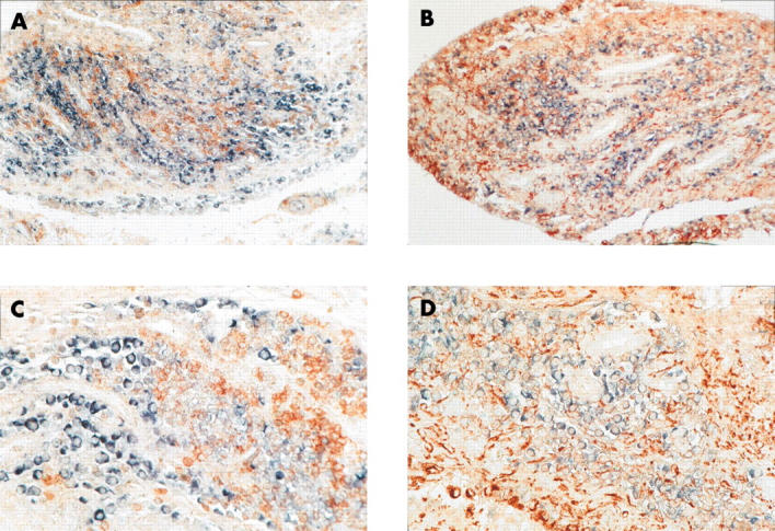
To further characterise the cell type expressing maspin, double labelling with anti-CD68 (A, C) and anti-CD3 antibodies (B, D) was performed after in situ hybridisation with maspin probes on paraffin embedded RA synovial tissue samples. Predominantly, maspin was detected in CD68 and CD3 negative SF. Blue-black colour: maspin transcripts, red-brown colour: anti-CD68 and anti-CD3 positive cells, respectively. Original magnificationx200 (A, C), x400 (B, D).