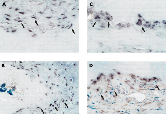
Figure 5.
Maspin mRNA expression was additionally investigated in RA synovial tissue at sites of cartilage and bone destruction. Maspin positive cells with both fibroblast-like (A) and macrophage-like as well as osteoclast-like morphology (C) were found. Double labelling with anti-CD68 antibodies showed both maspin positive, CD68 negative (figs 5B and 3G) as well as maspin positive, CD68 positive cells (D). Original magnification of all figures x400.

