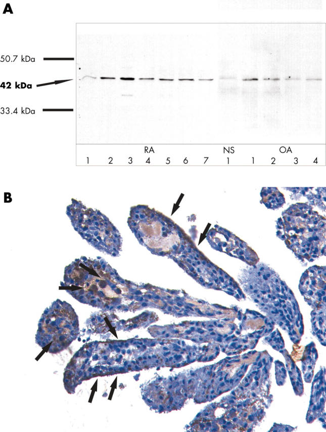
Figure 6.

To examine the protein expression of maspin in RA SF, SDS-PAGE and western blot with anti-maspin antibodies was performed in SF of seven patients with RA and four with OA. A positive band of the correct size (42 kDa) was detected in all RA SF and OA SF examined. In normal SF, a discrete band could also be detected (A). To confirm these results in situ, immunohistochemistry using anti-maspin antibodies was performed in synovial tissue samples of four patients with RA. Prostate tissue was used as positive control (result not shown). Maspin could be detected only in a few single cells in the synovial lining as well as single cells in the sublining (B, arrows). Original magnification x200.
