
Figure 1.
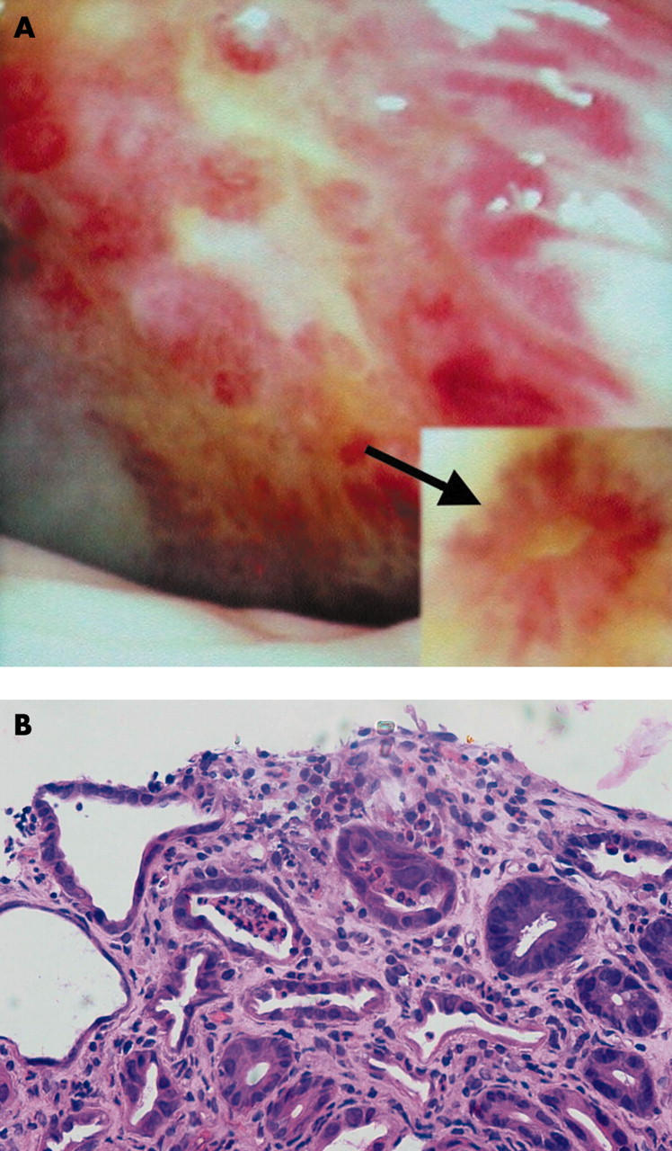
(A) The mucosa of the sigmoid colon was hyperaemic, oedematous, and markedly uneven, with multiple ulcers and haemorrhage. The ulcers were irregular in shape and varied in size. Some cotton wool-like vascular pattern (arrow) was also noted in the surrounding mucosa. (B) Colonic biopsy specimen showing inflammatory cell infiltrating the lamina propria, destruction of the mucosal gland, and marked crypt abscess (haematoxylin and eosin, x200)