
Figure 1.
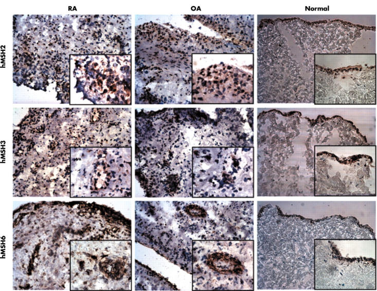
Distribution of DNA MMR enzymes in synovial tissue. Sections from RA (n = 6), OA (n = 5), and normal (n = 4) synovia were incubated with mAb to hMSH2, hMSH3, or hMSH6. The proteins were detected using the immunoperoxidase technique and DAB (brown colour). The slides were counterstained with haematoxylin (blue). Representative sections are shown. Images were obtained at x200 magnification, and each insert obtained at x400 magnification.