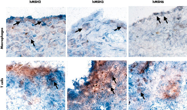
Figure 2.
Double label immunohistochemistry for MMR enzymes and cell surface markers. Representative field in an RA synovium are shown demonstrating the expression of hMSH2, hMSH3, and hMSH6 in CD3+ T cells and CD68+ macrophages. Examples of double positive cells are indicated by arrows. The most abundant staining for all three MMR proteins (red, peroxidase) was seen in intimal lining CD68+ (blue, AP) and CD68– cells (macrophage-like synoviocytes and fibroblast-like synoviocytes, respectively). Only rare T cells (blue, AP) in the sublining were positive (an example is shown in the figure).

