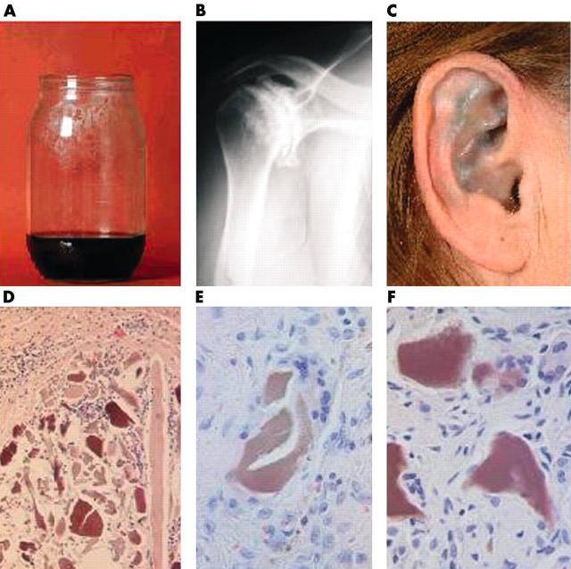
Figure 1 .
(A) Darkening of the urine by standing; (B) x ray picture of the right shoulder demonstrating a destroyed joint with sclerosis, osteophytosis; (C) slate-blue discolouration of the antihelix of the ear; (D) yellow-brown shards, scattered over the synovium; (E, F) shard evoking a foreign body reaction with multinucleated giant cells and surrounded by pigment loaded macrophages.

