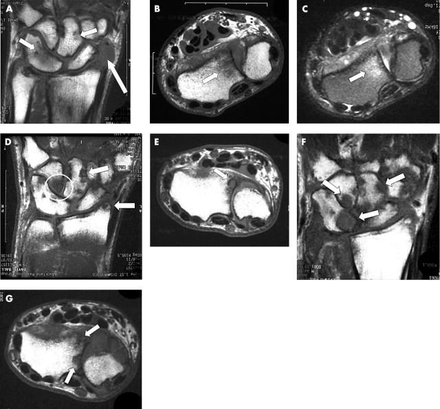
Figure 2 .
Sequential MR scans of the dominant carpus over 6 years in a 44 year old patient who was non-compliant with disease modifying treatment. This patient has congenital fusion of the triquetrum and lunate. (A) The baseline T1 weighted coronal scan shows focal areas of bone oedema as low signal within the capitate and the triquetrum/lunate (short arrows). There is extensive synovitis adjacent to the scaphoid (long arrow). (B) Axial T1 weighted image (baseline) showing bone oedema as low signal within the distal radius. (C) Equivalent baseline axial T2 weighted image showing bone oedema within the radius as bright signal. (D) Coronal T1 weighted scan at 1 year showing discrete low signal regions breaching the cortex of the capitate and the radial styloid indicating erosions (arrows). There is also extensive bone oedema within the hamate (circle). (E) Axial T1 weighted image at 1 year showing erosion at the radius. (F) Coronal T1 weighted scan at 6 years showing extensive erosive disease at multiple sites (arrows). (G) Axial T1 weighted image at 6 years showing more extensive erosion at the radius.

