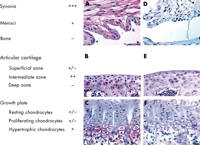
Figure 2 .
Recombinant adenoviruses infect different tissues and cells in the knee joint. Immunohistochemical localisation of ß-galactosidase in mouse knee joints using a monoclonal antibody was performed 1 week after injection with either RAdLacZ (A, B, and C) or a control virus RAd66 (D, E, and F). Representative sections of synovium (A and D), articular cartilage (B and E), and growth plate (C and F) are shown. These immunohistochemical analyses were also used to measure the transduction efficiency of RAdLacZ to various cells and tissues of the knee joint. Scoring shown to the left is based on the percentage of the cells positive for ß-galactosidase: –, no positive cells; +/–, 1–24%; +, 25–49%; ++, 50–74%; +++, 75–100%.

