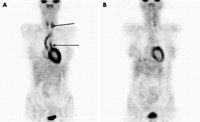
Figure 1.
(A) [18F]FDG-PET scan of patient 5 with active TA at diagnosis. Note the markedly abnormal uptake of [18F]FDG in the aortic arch and carotid arteries (arrows). (B) [18F]FDG-PET scan of the same patient in remission after treatment with prednisolone and intravenous cyclophosphamide. Note almost complete resolution of abnormal [18F]FDG uptake in these areas.

