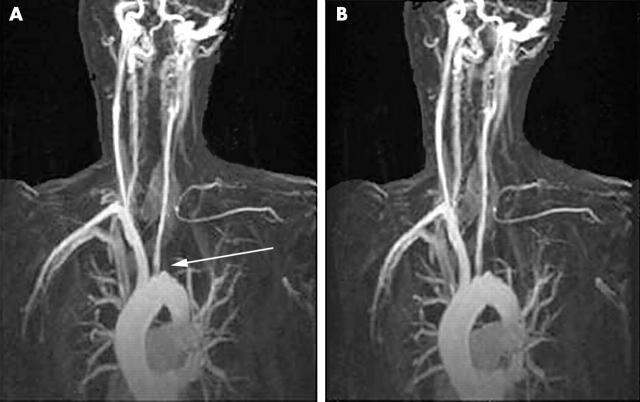
Figure 2.
(A) Magnetic resonance angiography (MRA) image from patient 5 with active TA at diagnosis. There is complete occlusion of the left subclavian artery at its origin (arrow) with numerous collaterals evident and an ostial stenosis of the left common carotid artery. (B) MRA image from the same patient in remission. No significant progression of the lesions found on the baseline MRA is seen.

