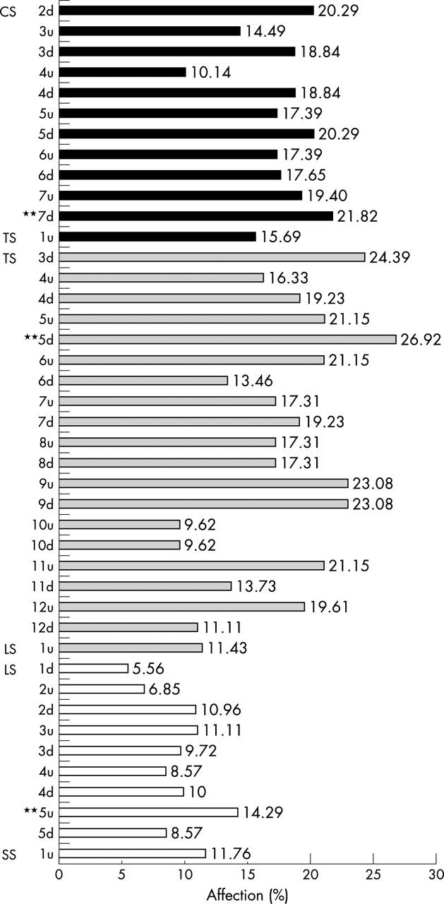
Figure 6.

Relative affection of each VU, as seen by x ray examination using the SASS score for evaluation of chronic spinal changes in patients with AS. Values are shown as the percentage of affection. u, upper edge of the VU; d, lower edge of the VU; SS, sacral spine. **VU with the most frequent affection in each spinal segment.
