
Figure 3.
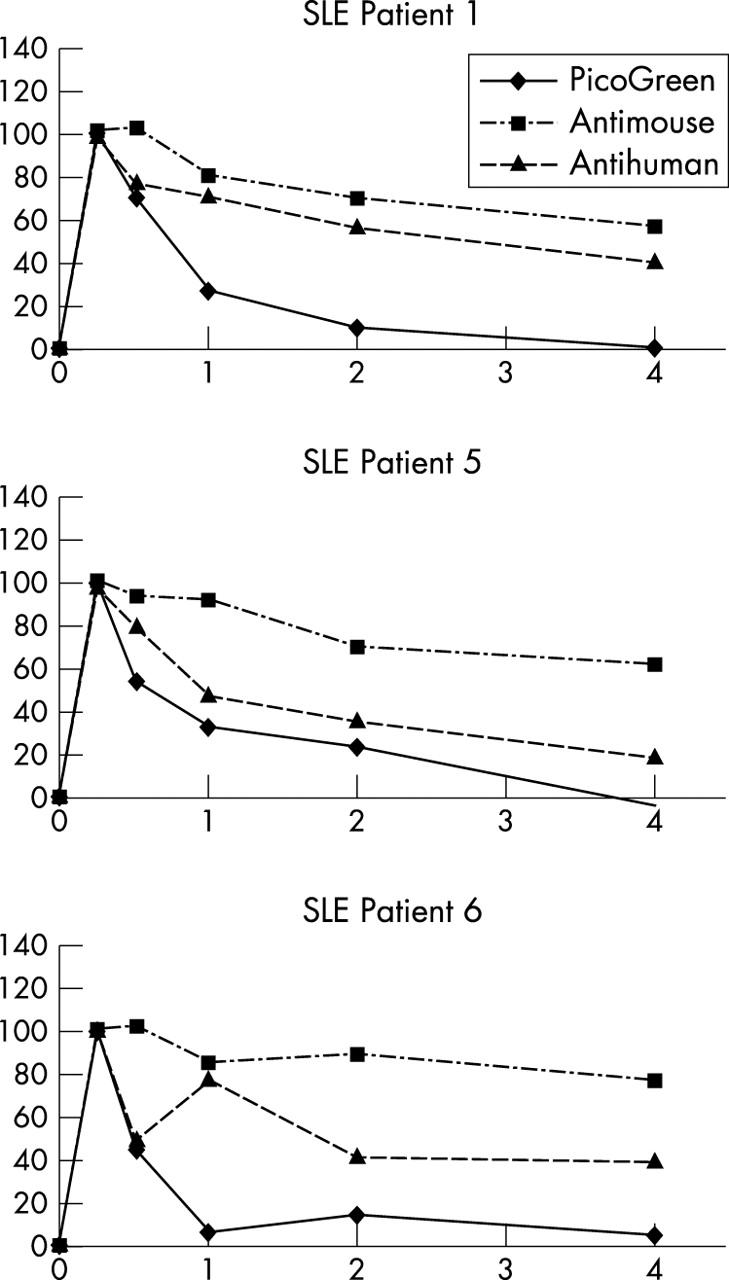
FACS analysis establishes binding of ETI-104 and target anti-dsDNA antibodies to RBCs in patients with SLE in vivo. RBCs from patients were stained after drug infusion to detect binding of the murine mAb (antimouse) and DNA (PicoGreen) components of ETI-104 and binding of the target anti-dsDNA antibody (antihuman). Average of the geometric mean fluorescence intensity of stained samples, after subtracting non-specific fluorescence, at each time was normalised to the 15 minute post-infusion value, which was set at 100%. The patients shown here represent the three patterns of association of anti-dsDNA antibodies with the RBCs; anti-dsDNA antibody tracked with the murine mAb component of ETI-104 (patient 1), with the DNA component of ETI-104 (patient 5), or non-parallel to either ETI-104 component (patient 6). Patients 2 and 3 showed essentially the same pattern as patient 1 and patient 4 as patient 5 (not shown). All values were measured in duplicate.