
Figure 5.
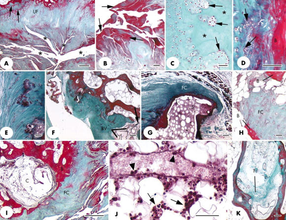
(A) Prominent fissures (*) at the ligament end of the medial epicondylar enthesis that greatly disrupt the normal structure of the attachment site. UF, uncalcified fibrocartilage; arrows, tidemarks. Scale bar = 500 µm. (B) Higher power view of several fissures from the same specimen as illustrated in (A) to show the prominent layer of fibrin (arrows) on the surface of the fissures. Scale bar = 50 µm. (C) Clusters of fibrocartilage cells (arrows) separated by small regions of acellular matrix (asterisk) in the tendon part of a lateral epicondylar enthesis. Scale bar = 50 µm. (D) Fibrocartilage cell proliferation (arrows) at the tendon attachment on the lateral epicondylar enthesis. B, bone. Scale bar = 100 µm. (E) Small foci of calcified tissue and fibrocartilage cell clusters in the zone of uncalcified fibrocartilage at the medial epicondylar enthesis. Scale bar = 50 µm. (F) Numerous small blood vessels (BV), indicating vascular proliferation at the site of a small bony defect (arrow) in the tendon attachment on the medial epicondylar enthesis. The blood vessels are passing between the tendon and the bone marrow. Scale bar = 100 µm. (G) A section adjacent to that featured in fig 2A to show the presence of fibrocartilage (FC) at the tip of the small bony spur (S). (H) A small region of fibrocartilage (FC) within the bone marrow, between neighbouring bone spicules (B). Scale bar = 50 µm. (I) A subchondral bone cyst (C) beneath the tendinous part of the lateral epicondylar enthesis. B, bone; FC, fibrocartilage. Scale bar = 500 µm. (J) A venule in the bone marrow associated with the tendinous part of the lateral epicondylar enthesis. Note the presence of inflammatory cells around the vessel (arrows) and of several marginating leucocytes around the edge of the lumen (arrowheads). Scale bar = 50 µm. (K) Evidence of bone marrow fibrosis (F) in the same specimen as (J). B, bone. Scale bar = 100 µm. All sections were stained with Masson's trichrome.