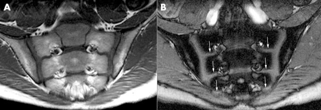
Figure 1.
October 1996 (age 13 years and 4 months). (A) T1 weighted fast spin echo sequence depicts smoothly marginated sacroiliac joints of normal width. (B) Contrast enhanced T1 weighted gradient echo sequence without enhancement related to sacroiliitis. Of note, the growth plates of the sacral bone (arrows) are seen.

