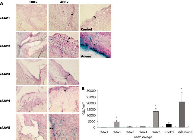
Figure 1.
ß-Gal expression in rat synovial tissue 2 weeks after intra-articular injection of different rAAV serotypes. Joints were snap frozen and sections were cut and stained in situ for ß-Gal activity and counterstained with nuclear red. Representative cryosections of right ankle joints of rats with adjuvant induced arthritis (AIA), injected with 6.1x10¹⁰ GC rAAV, Ad.LacZ as a positive control or saline as a negative control are shown (magnification x100, x400). The lining layer is indicated by arrows (A). Tissue sections were quantified for ß-Gal expression by computer assisted digital image analysis (B). Values are expressed as mean (SD) cumulative IOD/mm2. *p<0.05 as compared with the control group, Mann-Whitney U test.

