
Figure 2.
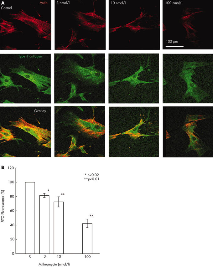
Evaluation of type I collagen production in systemic sclerosis dermal fibroblast monolayer cultures by confocal imaging after mithramycin treatment. Type I collagen present in individual cells of systemic sclerosis fibroblast cultures was examined by immunomicroscopy and confocal microscopy imaging as described in "Materials and methods" in five separate experiments using four cell lines. (A) Fluorescence of the FITC conjugated antirabbit IgG labelling type I collagen is shown in green and that of rhodamine labelled phalloidin labelling actin in red in one illustrative experiment. (B) Quantitative evaluation of the mithramycin effect on type I collagen production at the individual cell level showed that 48 hours of treatment with mithramycin at 10 nmol/l and 100 nmol/l decreased the protein level by about 33% and 50%, respectively. The results show the averages (SD) from five separate experiments with four different cell lines.