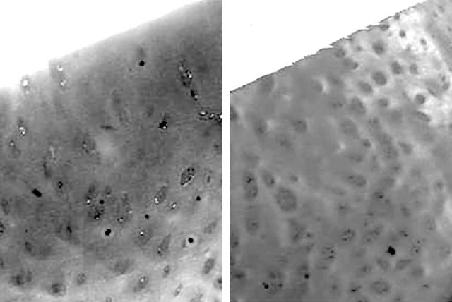
Figure 1.
Photomicrographs of safranin O stained histological sections of zones with macroscopically defined minimum (left) and maximum (right) damage in typical OA cartilage (original magnification x60). A diffuse staining of the cartilage, almost entirely masking the chondrocytes, is present in the minimum zone, while in the maximum zone there is evident hypocellularity together with some clusters of chondrocytes and severe reduction of the safranin O staining.

