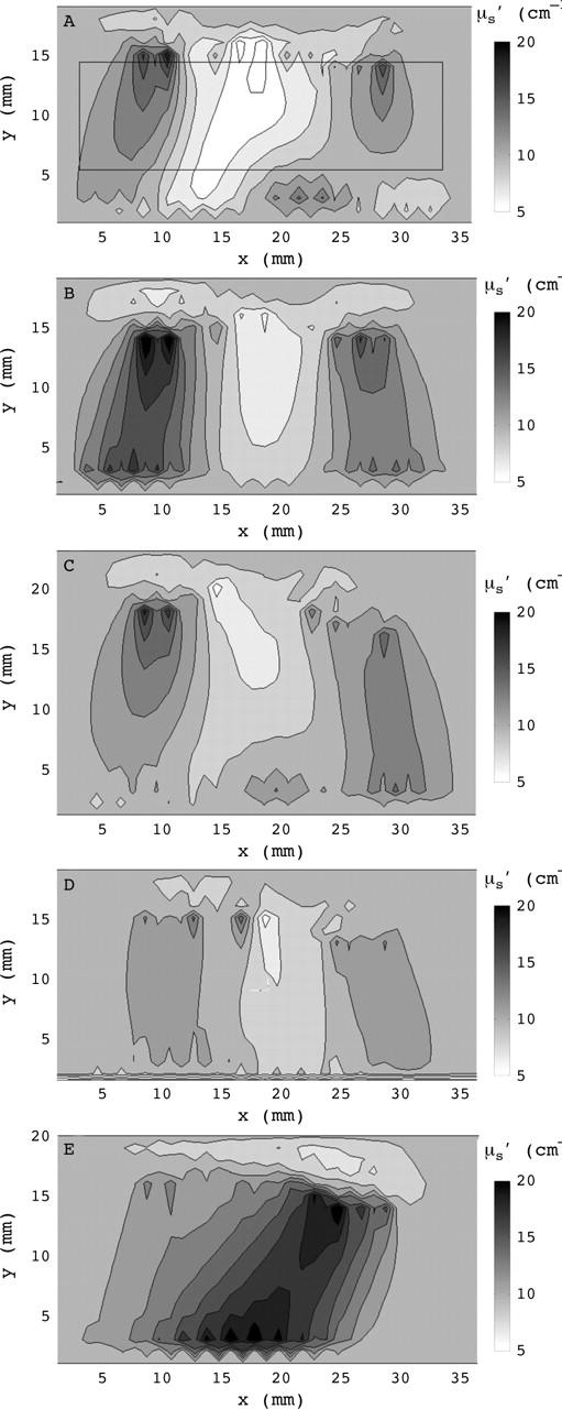
Figure 3.

Reconstructed cross sections of the scattering coefficient for three different fingers typical for (A) category 1—definitely no synovitis; (B) category 2—probably no synovitis; (C) category 3—possibly synovitis; (D) category 4—probably synovitis; and (E) category 5—definitely synovitis. The fingertip is located to the right of the images that show a 36 mm wide section of the finger with the joint cavity located approximately in the centre. The rectangle in fig 3A indicates the region for which Min(µs), Min(µa), Max(µs), Max(µa), Min(µs)/Max(µs), and Min(µa)/Max(µa), were calculated.
