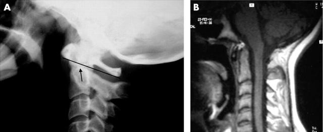
Figure 2.
(A) Lateral view radiograph of case 2. No AAI is present according to the Sakaguchi-Kauppi (S-K) method,6 which divides the AAI phenomenon into four grades. In lateral view radiographs of the cervical spine, the upper lateral facets of the axis form an easily visible "curve" on the side of the axis (the pedicle). In normal cases (grade I) as in this case, "the cranial tip of this curve" (arrow) is situated caudally from the line, drawn between the most caudal parts of the anterior and posterior arch of the atlas (lower atlas line; drawn in the figure). Owing to erosions of the facets, the atlas may fall down around the axis and the "tip" may reach or penetrate the "lower atlas line" and AAI is then diagnosed (grade II). Grade III means that "the tip of the curve" is on the level (or above) of the line drawn between the central points of the atlas arches. Grade IV means that a very severe AAI is present, with collapsed lateral facets, and the "tip" is on the level (or above) of the line drawn between the most cranial parts of the atlas arches.6 (B) MRI of the cervical spine of case 2. The spinal canal is normal and there is no compression of the spinal cord. Active synovitis may be present above the dens of the axis.

