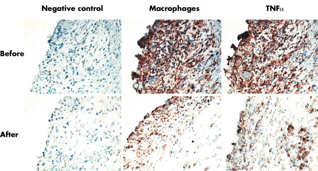
Figure 2.
Pathological specimen of pigmented villonodular synovitis before and after 20 weeks of treatment with anti-TNFα. Before treatment: classic histological features were seen, with pigment depositions and an impressive infiltrate, mainly consisting of macrophages with an abundant presence of TNFα, and immunohistochemical staining for macrophages and TNFα at baseline. After treatment: a dramatic reduction in cellularity, number of macrophages, estimated pigment, and TNFα after 20 weeks of treatment with anti-TNFα was seen.

