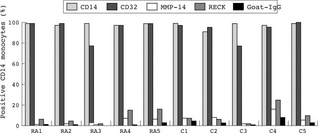
Figure 7.
Expression of RECK, MMP-14, CD14, and CD32 (FcγRII) on monocytes from five patients with RA and five healthy controls as determined by flow cytometric analysis. Monocytes were detected in sodium citrate treated blood. The percentage of positive cells was determined. Data were evaluated by the Mann-Whitney U test (p<0.05). Note that only low levels of cells express RECK and MMP-14. No significant differences in RECK expression on monocytes was found between patients with RA and healthy controls. RA, rheumatoid arthritis; C, control

