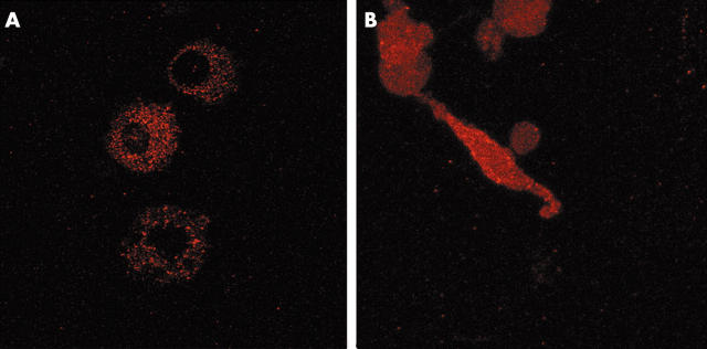
Figure 8.
Immunolocalisation of RECK in RA monocytes treated for 7 days with M-CSF and fibroblasts isolated from RA synovial membranes, and subsequently cultured for 3 days. Confocal microscopy shows that in macrophages, RECK is only expressed intracellularly and not on its membrane (A). In contrast, synovial fibroblasts showed a dispersed pattern and RECK was clearly present on the membrane (B). Original magnification x400.

