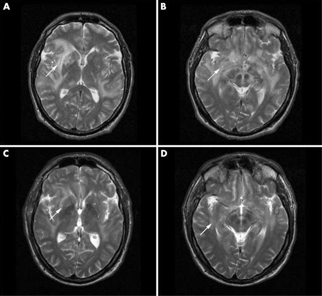
Figure 1.
(A, B) Axial T2 weighted fast spin echo MR images of the brain show increased signal intensity in the bilateral white matter and basal ganglia (arrows). (C, D) Follow up MRI shows a dramatic improvement of the white matter and basal ganglia abnormalities only 8 weeks after starting treatment.

