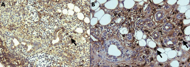
Figure 1.
(A) Immunohistochemical staining for IgA positive plasma cells in parotid gland biopsy specimen before treatment, showing a few IgA positive plasma cells (arrow) and a massive infiltrate with a few ducts (magnification x200). (B) Immunohistochemical staining for IgA positive plasma cells in parotid gland biopsy specimen after rituximab treatment, showing less infiltrate and more salivary gland ducts, with a relative increase of IgA positive plasma cells (arrows) (magnification x200).

