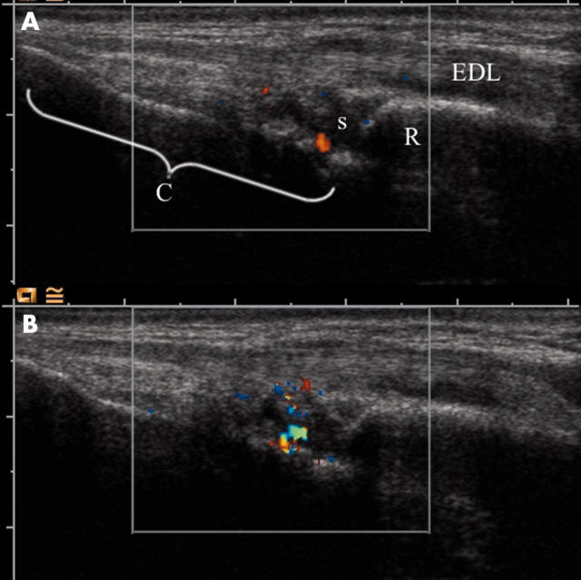
Figure 1.
Colour Doppler in the wrist before and after SonoVue injection. The images are longitudinal through the extensor digitorum longus tendon (EDL). The surface of the radius (R) and carpal bones (C) are seen as bright reflectors. The synovium of the radiocarpal joint(s) is seen as a anechoic/hypoechoic mass with extensions that are synovial duplications. (A) Before contrast injection. A single Doppler focus is visible inside the synovium. (B) After contrast injection. A larger Doppler focus is visible.

