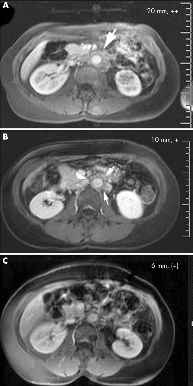
Figure 1.

Serial MRI study of RPF during immunosuppressive treatment. T1 weighted images of patient 2 after (A) 2, (B) 14, and (C) 26 months of immunosuppression, indicating the diameter of the periaortic mantle and gadolinium enhancement (absent "–", detectable "(+)", intermediate "+" and strong "++").
