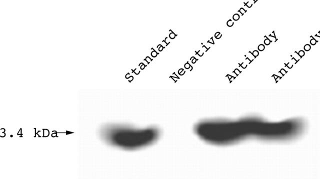
Figure 4.
Expression of humanin peptide in synovial cells from diffuse-type PVNS. Protein (20 µg) from synovial cell lysates was subjected to SDS-PAGE on a 5–20% gradient gel. Rabbit anti-humanin polyclonal antibody was used for western blotting. Synthesised peptide, which was used as antigen to produce rabbit anti-humanin polyclonal antibody, was used as a standard and rabbit IgG was used as a negative control.

