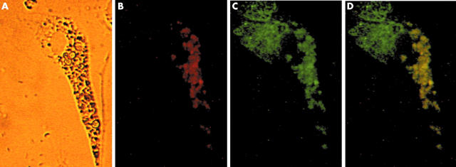
Figure 6.
Relationship between humanin peptide expression and mitochondria. Isolated synovial cells containing haemosiderin were double stained with anti-humanin antibody and anti hsp60 antibody as first antibodies, followed by goat antirabbit IgG and donkey antigoat IgG as second antibodies (x400). (A) Haemosiderin was deposited unequally throughout the cytoplasm. (B) Single anti-humanin antibody staining (red). (C) Single anti-hsp60 antibody staining (mitochondrial staining; green). (D) Humanin was dominantly distributed in the mitochondria around the siderosome (yellow).

