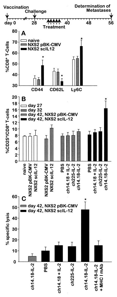
Figure 1.
Effect of ch14.18-IL-2 treatment on reactivation of CD8+ T cells after initial vaccination with scIL-12 NXS2 cells. (A) Phenotype of CD8+ T cells after vaccination of A/J mice with NXS2 cells genetically engineered to secrete scIL-12. Splenocytes of mice (n = 4) injected with scIL-12 NXS2, NXS2 cells carrying the empty vector, or naive mice were analyzed 28 days after vaccination by two-color flow cytometry. Differences between scIL-12 NXS2 vaccinated and naive mice or mice receiving the irrelevant vaccine were statistically signif icant. ∗, P < 0.02. (B) Detection of activated CD8+ T cells after vaccination, challenge, and booster injection with ch14.18-IL-2 fusion protein. Splenocytes of naive mice (n = 4), scIL-12 NXS2-vaccinated mice, and mice injected with NXS2 cells carrying the empty vector were analyzed for CD25+/CD8+ T cells 5 days after five daily i.v. injections of 10 μg of ch14.18-IL-2, an equivalent mixture of 10 μg of ch14.18 antibody and 30,000 units of IL-2, or 10 μg of nonspecific ch225-IL-2 fusion protein. Differences among scIL-12 NXS2-vaccinated mice and mice receiving a ch14.18-IL-2 booster injection, and all control groups was statistically significant. ∗, P < 0.01. (C) Determination of CD8+ T cell-mediated cytotoxicity after vaccination and booster injections with ch14.18-IL-2 fusion protein. CD8+ T cells of scIL-12 NXS2-vaccinated mice (n = 4) and mice injected with NXS2 cells carrying the empty vector were isolated 5 days after the booster injections as described in B and used in a standard 51Cr release assay with NXS2 target cells. Differences among scIL-12 NXS2-vaccinated mice and mice receiving ch14.18-IL-2 booster injections, and all control groups was statistically significant. ∗, P < 0.001.

