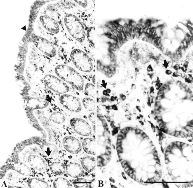
Figure 3 .

Biopsies from a healthy large intestine processed to demonstrate immunoreactivity of antisecretory factor (AF). Bars=100 µm. (A) Low magnification showing AF immunoreactivity in the surface epithelium (arrowhead), in crypt cells, and in lymphocyte-like cells (arrows) in the lamina propria. (B) Larger magnification showing the AF positive lymphocyte-like cells (arrows).
