Figure 1.
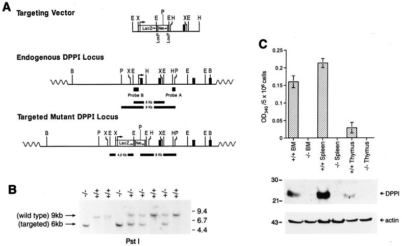
Targeting of the DPPI gene. (A) Creation of the targeting vector. The DPPI locus is shown in the Middle and the targeting construct is depicted at Top. The locations and transcriptional orientations of the β-galactosidase (LacZ) and PGK-Neo cassettes are indicated. The targeting construct was designed to replace exon 1 and part of intron 1 with the selectable marker cassette. The locations of the external (probe A) and internal (probe B) probes used to detect homologous recombination are shown. (B) Southern blot analysis of tail DNA from wild-type, DPPI+/−, and DPPI−/− animals. DNA was digested with PstI and hybridized with probe A. The wild-type allele is found within a 9-kb fragment; the targeted allele is reduced to 6 kb because of an internal PstI site in the PGK-Neo cassette. (C) DPPI activity in wild-type and DPPI−/− tissues. Freshly isolated bone marrow cells, splenocytes, and thymocytes were lysed and assayed for DPPI activity as determined by the hydrolysis of Gly-Phe-β-naphthylamide. Activity was defined as the OD at 405 nm for 5 × 10⁶ cells. Results represent the mean of duplicate determinations. Equal amounts of total lysates were also fractionated on 10% SDS/PAGE gels and immunoblotted with a specific rabbit anti-mouse DPPI antibody. A β-actin antibody was used to control for protein content and loading.

