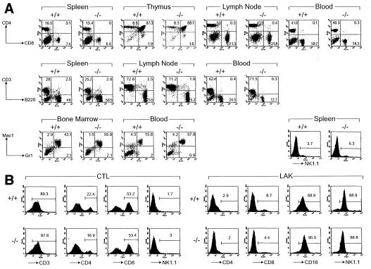
Figure 2.
Normal immune system development and cytotoxic lymphocyte activation in DPPI mutant mice. (A) Flow cytometric analyses of lymphoid and myeloid cells in wild-type and DPPI−/− mice. Cells harvested from spleen, thymus, mesenteric lymph nodes, and peripheral blood were analyzed for the expression of the indicated surface markers. The percentages of cells expressing CD3, CD4, CD8, B220, and NK1.1 were similar for all mice. Note that there was a moderate increase in the Mac-1+/Gr-1+ in the example shown here; 3 of 11 tested mice had a similar finding. (B) Normal proliferation and activation of DPPI−/− splenocytes in primary MLR and LAK activation. Splenocytes from wild-type and DPPI−/− animals were activated in a 5-day mixed lymphocyte reaction culture or in the presence of high-dose IL-2 to generate LAK cells. The expansion of the CD8+ CTL and NK1.1+ LAK cell populations were essentially equivalent in DPPI+/+ and −/− splenocytes.

