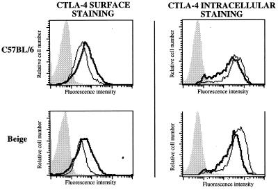
Figure 6.
CTLA-4 surface and intracellular expression of pervanadate-treated T cells from C57BL/6 and beige mice. T cells isolated from the lymph nodes of C57BL/6 and beige mice were stained for surface and intracellular CTLA-4 after being activated for 96 h with γ-irradiated (2000 rads) spleen cells prepared from DBA/2 mice. Cells were incubated with (thick lines) or without (thin lines) pervanadate.

