
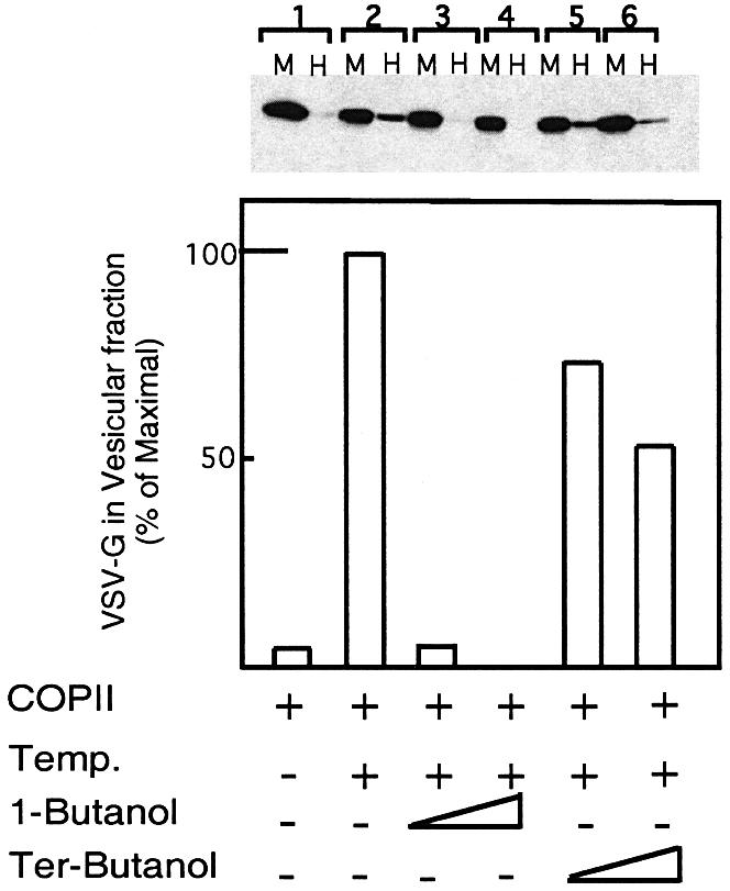
Fig. 2. PLD activity is required for COPII-mediated export from the ER. VSV-G-containing membranes were incubated with purified COPII components [Sar1p-H79G (2 µg) Sec23/24 (1 µg) and Sec13/31 (12 µg)] (Aridor et al., 1998) for 30 min on ice (lane 1), or at 32°C (lanes 2–6) in the absence (lanes 1 and 2) or presence of 1-butanol (lane 3, 1%; lane 4, 2%) or tert-butanol (lane 5, 1%; lane 6, 2%). At the end of the incubation, the vesicle fraction (H for high speed pellet) was separated from the donor membranes (M for medium speed pellet) by differential centrifugation, and the export of VSV-G into the vesicular fraction was determined by western blot analysis with anti-VSV-G antibody. The lower panel is a quantitative densitometry analysis of the amount of VSV-G in the vesicle fraction. Results are presented as a percentage of maximal budding under non-perturbed conditions (25% of total VSV-G in the starting membrane, lane 2). The experiment presented is representative of at least three independent experiments.