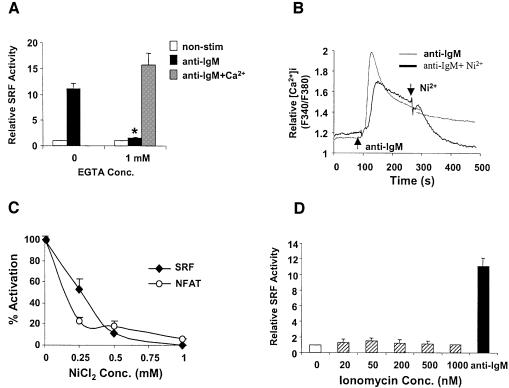
Fig. 3. Extracellular Ca2+ influx is essential but not sufficient for BCR-mediated SRF activation in WT DT40 cells. (A) BCR-mediated SRF activation is abrogated by chelating extracellular Ca2+ with EGTA. SRF transcriptional activity was determined in WT DT40 cells treated with or without anti-IgM in normal Ca2+ concentration, lacking extracellular Ca2+ (by adding EGTA) or in the presence of EGTA and reconstituted with Ca2+ (equimolar EGTA and Ca2+ concentrations). *P < 0.05 versus control without EGTA. (B) NiCl2 blocks BCR-induced intracellular Ca2+ elevation. Intracellular Ca2+ response induced by anti-IgM stimulation with (bold line) or without (thin line) 1 mM NiCl2 added at the indicated time in WT DT40 cells. The results are representative of three independent experiments. (C) NiCl2 inhibits BCR-induced SRF activation. SRF transcriptional activity was determined in WT DT40 cells stimulated with anti-IgM in the absence or presence of the indicated concentrations of NiCl2. The SRF–luciferase activity is shown as % activation (0 and 100% activation refer to no stimulation or stimulation with anti-IgM in normal medium, respectively, and are the mean ± SD of two independent experiments each performed in triplicate). (D) Intracellular Ca2+ elevation is not sufficient for SRF activation. SRF transcriptional activity was determined in WT DT40 cells treated with the indicated concentrations of ionomycin before harvesting for luciferase assay.

An official website of the United States government
Here's how you know
Official websites use .gov
A
.gov website belongs to an official
government organization in the United States.
Secure .gov websites use HTTPS
A lock (
) or https:// means you've safely
connected to the .gov website. Share sensitive
information only on official, secure websites.
