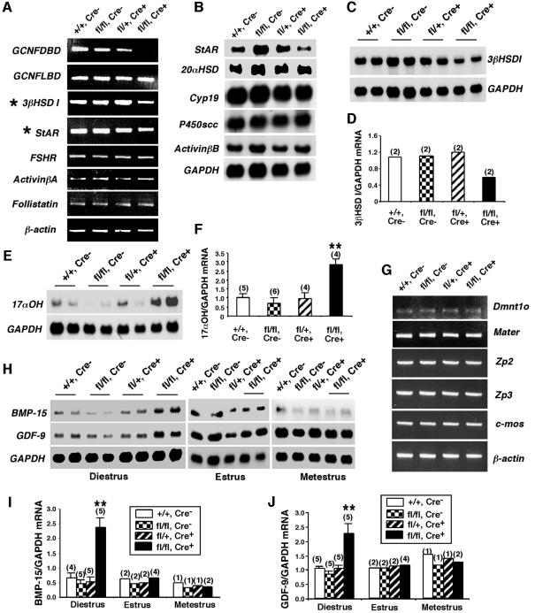
Fig. 4. Expression of ovarian marker genes in the GCNFfl/flZp3Cre+ ovary. (A–F) Expression of key ovarian genes expressed in somatic cells of GCNF+/+ (+/+, Cre–), GCNFfl/fl (fl/fl, Cre–), GCNFfl/+Zp3Cre+ (fl/+, Cre+) and GCNFfl/flZp3Cre+ (fl/fl, Cre+) ovaries at diestrus (day 1–2). (A) RT–PCR showing the reduced StAR and 3βHSD I expression in the GCNFfl/flZp3Cre+ mice. (B–F) Nothern blot analyses showing the mis-expression of (B) StAR, (C and D) 3βHSD I and (E and F) 17αOH in GCNFfl/flZp3Cre+ mice. Quantitative 3βHSD I and 17αOH mRNA levels in the northern blots are shown in the bar graph in (D) and (F), respectively. (G–J) Expression of oocyte marker genes. (G) RT–PCR showing expression of oocyte genes, Dmnt1o, Mater, Zp2, Zp3 and c-mos, in ovaries. Experiments were repeated twice using two individual animals. (H) Representative radiographs of northern blot analyses showing the BMP-15 and GDF-9 expression. (I and J) Quantitative ovarian (I) BMP-15 and (J) GDF-9 mRNA levels in northern blots. For (F), (I) and (J), relative mRNA levels (normalized to GAPDH mRNA levels) are presented as means ± SEs from various numbers of animal samples (indicated by alphabetic numbers) of each genotype. **P < 0.01 compared with three other groups at diestrus.

An official website of the United States government
Here's how you know
Official websites use .gov
A
.gov website belongs to an official
government organization in the United States.
Secure .gov websites use HTTPS
A lock (
) or https:// means you've safely
connected to the .gov website. Share sensitive
information only on official, secure websites.
