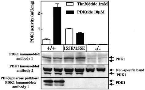
Fig. 2. Expression and activity of PDK1 in knock-in ES cells. The indicated ES cells were cultured to 80% confluence and lysed. PDK1 was immunoprecipitated from the cell lysate and assayed with the indicated peptide as described in Materials and methods. The results shown are the average ± SEM of three separate dishes of cells with each assay performed in duplicate. The cell lysates were also immunoblotted with PDK1 antibody 1 (raised against the C-terminal 20 residues of mouse PDK1) or PDK1 antibody 2 (raised against recombinant human PDK1 protein). The lysates were also incubated with Sepharose conjugated to PIF to affinity purify PDK1 as described in Materials and methods. The washed resin was then immunoblotted for PDK1 using PDK1 antibody 1. Similar results were obtained in two separate experiments. It should be noted that PDK1 in ES cells, as observed in other cell lines, is detected as two bands on immunoblot analysis (Balendran et al., 1999a; Williams et al., 2000).

An official website of the United States government
Here's how you know
Official websites use .gov
A
.gov website belongs to an official
government organization in the United States.
Secure .gov websites use HTTPS
A lock (
) or https:// means you've safely
connected to the .gov website. Share sensitive
information only on official, secure websites.
