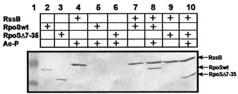
Fig. 4. σS(Δ7–35) binds to phosphorylated RssB in vitro. Equimolar amounts (0.3 nmol) of S-TRX-His6-RssB and His6-σS or His6-σS(Δ7–35) were incubated at room temperature with or without 50 mM acetyl phosphate (Ac-P), and subject to affinity chromatography on S-protein agarose, separated by SDS–PAGE and visualized using Penta-His antibodies. Size standard proteins (49 and 32.5 kDa) are shown in lane 1. In lanes 2 and 3, His6-σS and His6-σS(Δ7–35), respectively, were directly loaded on the gel.

An official website of the United States government
Here's how you know
Official websites use .gov
A
.gov website belongs to an official
government organization in the United States.
Secure .gov websites use HTTPS
A lock (
) or https:// means you've safely
connected to the .gov website. Share sensitive
information only on official, secure websites.
