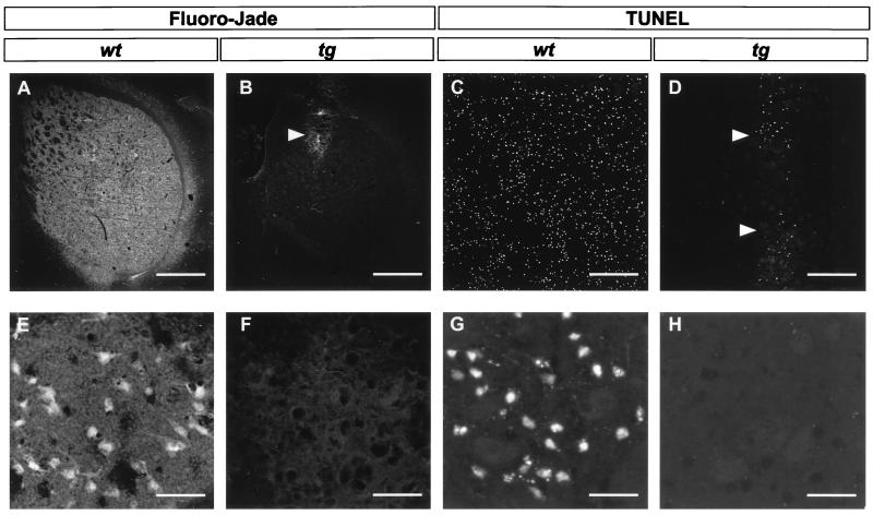
Figure 1.
Micrographs of striatal sections prepared from brains 48 h after intrastriatal quinolinic acid injection, labeled with the two fluorescent cell death markers Fluoro-Jade (A, B, E, and F) or TUNEL (C, D, G, and H). Sections from wild-type mice (wt; A, C, E, and G) contain numerous stained cells. In sections from transgenic mice (tg; B, D, F, and H) only very few cells are labeled along the cannula track (arrowheads). [Bar = 720 μm (A and B), 250 μm (C and D), and 30 μm (E–H).]

