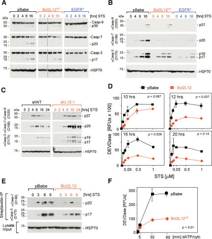
Figure 3.
Bcl2L12 inhibits post-mitochondrial effector caspase activity. (A,B) Ink4a/Arf-deficient astrocytic cell cultures ectopically expressing pBabe, Bcl2L12V5, and EGFR* were treated with STS (1 μM) for the indicated periods of time and were subjected to Western blot analyses using antibodies specific for the procaspases (A) and active caspases (B). For A, images from two minigels that were blotted onto the same membrane were pasted together: Samples 0, 2, 4, 8, and 16 (pBabe), and 0 and 2 (Bcl2L12) were loaded on one gel, and samples 4, 8, and 16 (Bcl2L12), and 0, 2, 4, 8, and 16 (EGFR*) were loaded on a second gel. We added a dashed line to indicate this. Comparable results were obtained using a STS concentration of 100 nM (data not shown). (C) U87MG cells stably expressing a nontargeting control (shNT) or a Bcl2L12-specific shRNA (shL12-1) were treated with STS (1 μM) for the indicated periods of time, lysed, and subjected to Western blot analysis as in B. (D) Ink4a/Arf-deficient astrocytic cell cultures ectopically expressing pBabe or Bcl2L12V5 were treated with the indicated doses of STS and were assayed for DEVDase activity at 10, 12, 16, and 20 h using an AFC-labeled DEVD peptide. P values were calculated for the 1 μM doses. (E) pBabe- and Bcl2L12-expressing astrocytes were treated with STS (1 μM) for the indicated periods of time and lysed, and active caspases were affinity-labeled with biotinylated VAD-fmk. Streptavidin precipitates were analyzed by Western blot using antibodies specific for active caspase-3 and caspase-7. (F) Lysates from pBabe- and Bcl2L12-expressing astrocyte cultures were activated with dATP (1 mM) and cytochrome c (5 μM), and DEVDase activity was monitored using AFC-labeled DEVD peptide. The p value was calculated for the 60-min time point. Error bars represent standard deviations of replicate data points, and two-tailed p values were calculated using the Student’s t-test.

