
Figure 3.
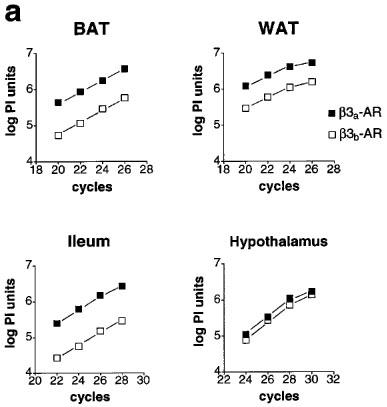
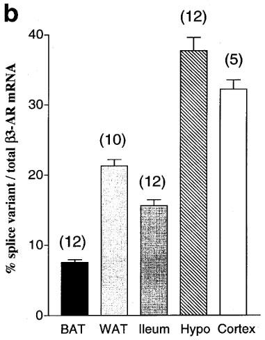
Relative levels of β3a-AR and β3b-AR mRNA. (a) Product/cycle relationship for β3-AR PCR performed on cDNA from BAT, WAT, ileum and hypothalamus. The cDNA was produced by reverse transcription of 1 μg total RNA, and one tenth used for PCR with β3-AR primers (mb3.f1 and mb3.r1). For each tissue, a single PCR mix was aliquotted into four separate capillaries which were removed sequentially from the cycler and placed on ice after the cycle numbers shown. Following gel electrophoresis and transfer to Hybond N+, PCR products were detected by hybridization with a [33P]-labelled β3-AR probe (mb3.pA) and exposure to phosphorimager plates for 16 h. The β3a-AR and β3b-AR products were quantitated using ImageQuaNT software (Molecular Dynamics), and the log of the volume (phosphorimager units, PI) plotted against cycle number. Note that the lines representing accumulation of the β3a-AR and β3b-AR PCR products are parallel, indicating proportional amplification. (b) β3b-AR splice variant mRNA as a percentage of total β3-AR mRNA in BAT, WAT, ileum, hypothalamus and cortex. Bars show the mean (s.e.mean). β3a-AR and β3b-AR fragments were quantitated as in (a) from multiple tissue samples (5 to 12).