Abstract
Cardiac calcinosis is a common complication of end stage renal disease. A newly observed risk of thromboembolism is reported in four patients with mobile cardiac calcinosis, treated with long term dialysis. Rapidly growing mobile calcification was confirmed by echocardiography. Each patient had an imbalance in serum calcium × inorganic phosphate (Ca × P product ⩾ 50); this imbalance could not be treated due to the sudden death of the patient or the need for surgical resection to prevent recurrent cerebral thromboembolism. Histological examination revealed intracardiac calcinosis in three cases, and each case showed haemodialysis hypoparathyroidism (intact PTH < 160 pg/ml). Thromboembolism in such cases is rare, however it indicates a need for cautious echocardiographic monitoring in end stage renal disease in patients with an uncontrolled Ca × P product. Keywords: cardiac mass; intracardiac mobile calcinosis; haemodialysis hypoparathyroidism; thromboembolism; end stage renal disease
Full Text
The Full Text of this article is available as a PDF (85.1 KB).
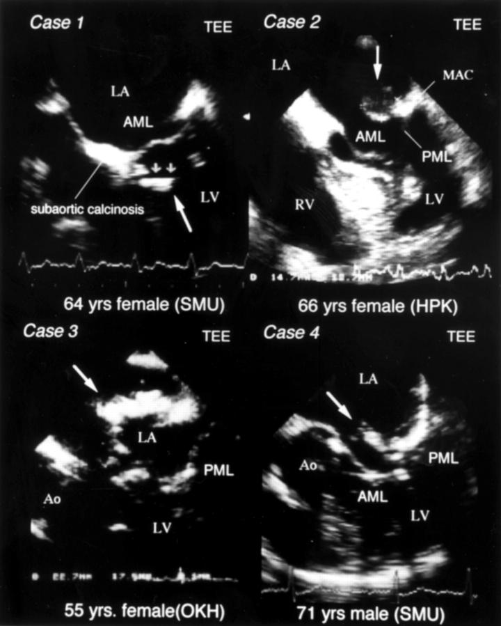
Figure 1 .
Case 1: transoesophageal echocardiography (TEE) showing a mobile intracardiac tumour (6 × 14 mm) with homogeneous hyperechoic characteristics, attached to the membranous portion of interventricular septum with tumour. Case 2: echocardiography after cerebral thromboembolism, showing a heterogenic high echoic mobile intracardiac tumour at the base of the posterior mitral leaflet (PML), and the rapid growth of the tumour (14.7 × 13.7 mm). Case 3: echocardiography showing rapidly growing intracardiac mass (14 × 12 mm) on posterior wall of left atrium (LA; mid-panel). Case 4: transoesophageal echocardiography showing a homogeneous high echoic mobile intra-atrial tumour located in anterior mitral valve (AMV) (8 × 12 mm). AML, anterior mitral leaflet; Ao, aorta; LV, left ventricle; MAC, mitral annular calcinosis; RV, right ventricle.


